1) right ventricular bifocal

右室双部位
1.
Objective: To assess recent clinical effect of right ventricular bifocal(RV-Bi) pacing with echocardiography.
目的:用超声心动图观察右室双部位(RV-B i)起搏的临床效果。
2) biatrial-right ventricular upper septal
双心房-右室间隔上部
3) Right ventricular apex
右室心尖部
1.
AIM To compare the effects of pacing on hemodynamics in right ventricular septum and in right ventricular apex.
目的比较右室流出道间隔部(RVS)与右室心尖部(RVA)起搏对血流动力学的影响。
2.
AIM:To compare the cardiac function between right ventricular septal pacing and right ventricular apex pacing.
结论右室室间隔起搏较右室心尖部起搏更利于起搏器植入患者的心功能保护。
4) right ventricular apical
右室心尖部
1.
The effect of the pacing between right ventricular outflow septum and right ventricular apical on the heart function.;
目的了解右室流出道间隔部起搏和右室心尖部起搏参数的差异及对心功能的影响。
5) Double-outlet right ventricle
右室双出口
1.
The clinic value of Surgical treatment for 35 cases of double-outlet right ventricle
手术治疗右室双出口35例临床分析
2.
Objective: To report the experiences of surgical treatment of tetralogy of Fallot(TOF) and double-outlet right ventricle(DORV) with anomalous coronary artery and explore the influence of coronary artery malformation for reconstructing outflow tract of right ventricle.
目的:总结法乐四联症和右室双出口合并冠状动脉畸形手术治疗经验,探讨冠状动脉畸形对右室流出道重建的影响。
3.
Objective In order to optimize the surgical indications and managements of double-outlet right ventricle(DORV)repaired with intraventricular tunnel, the case selection, anatomical features, surgical managements and results were reviewed.
方法 12 0例右室双出口 (DORV)患者接受心室内隧道修补手术 ,采用纵行切开右室流出道 ,用一补片作右心室内隧道 ,连接室间隔缺损与主动脉口 ,引导左心室血在补片下进入主动脉 ,根据解剖情况扩大右室流出道和肺动脉。
6) Double-chambered right ventricle
双腔右心室
1.
Surgical treatment of double-chambered right ventricle: a report of 95 cases;
双腔右心室95例的外科治疗
2.
Objective The value of echocardiography in the diagnosis of double-chambered right ventricle(DCRV) is analyzed.
目的 探讨超声心动图对诊断双腔右心室 (DCRV)的价值 ;方法 总结 1995年 1月~ 2 0 0 1年 5月间经手术证实为双腔右心室的 12例住院病例资料 ,分析其超声心动图特点 ,并与手术结果进行对照 ;结果 超声心动图上右心室腔内室上嵴至右心室游离壁见粗大肌束横跨 ,中央孔狭窄 ,将右心室分成高压腔及低压腔 ,彩色多普勒血流显像(CDFI)在中央孔示五色镶嵌的湍流 ,CW估测中央孔狭窄压差来判断病变程度 ;12例患者中 ,超声心动图诊断DCRV与手术符合 10例 ,符合率为 83 3% ;DCRV以合并室间隔畸形最为常见 ;结论 超声心动图在诊断DCRV及其合并心脏畸形等方面具可靠价值 ,已成为临床上术前正确诊断及术后疗效评价重要手
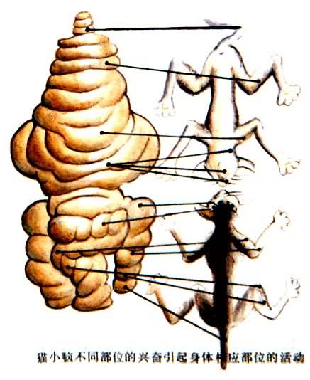
补充资料:人脑的功能分化猫小脑不同部位的兴奋引起身体相应部位的活动
李瑞端绘
[图]
说明:补充资料仅用于学习参考,请勿用于其它任何用途。
参考词条